Did you know that the colonoscope must navigate 160-180cm through your intestinal tract, while the gastroscope only travels 100-120cm to reach its destination? Colonoscopy examines your large intestine from rectum to cecum using a flexible tube with a camera, while gastroscopy investigates your upper digestive tract from mouth to duodenum. These endoscopic procedures serve distinct diagnostic purposes, require different preparation protocols, and detect separate sets of conditions.
Both procedures involve sedation, real-time visualization, and the ability to perform biopsies or remove polyps during the same session. Your gastroenterologist might recommend one or both procedures based on your symptoms—rectal bleeding typically prompts colonoscopy, while persistent heartburn or difficulty swallowing indicates gastroscopy. The choice between colonoscopy vs gastroscopy depends on whether your symptoms suggest lower or upper gastrointestinal issues.
BLOG ARTICLE
Anatomical Coverage and Procedure Specifics
Colonoscopy Examination Areas
The colonoscope enters through the rectum and navigates through the entire colon, examining the rectum, sigmoid colon, descending colon, transverse colon, ascending colon, and cecum. During withdrawal, the gastroenterologist systematically inspects the intestinal walls for polyps, inflammation, diverticula, or abnormal tissue. The procedure typically takes 20-45 minutes, with additional time needed if polyps require removal.
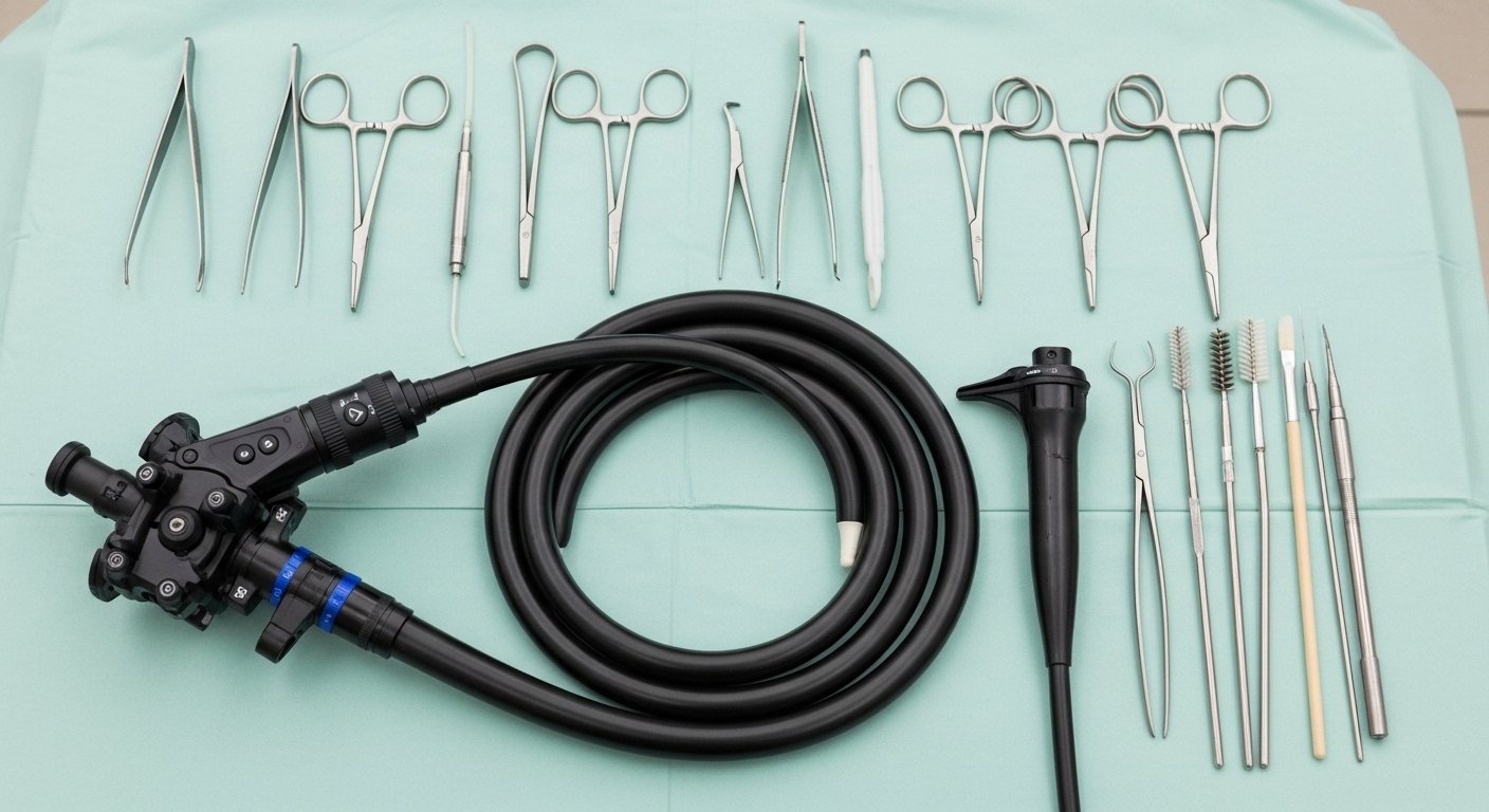
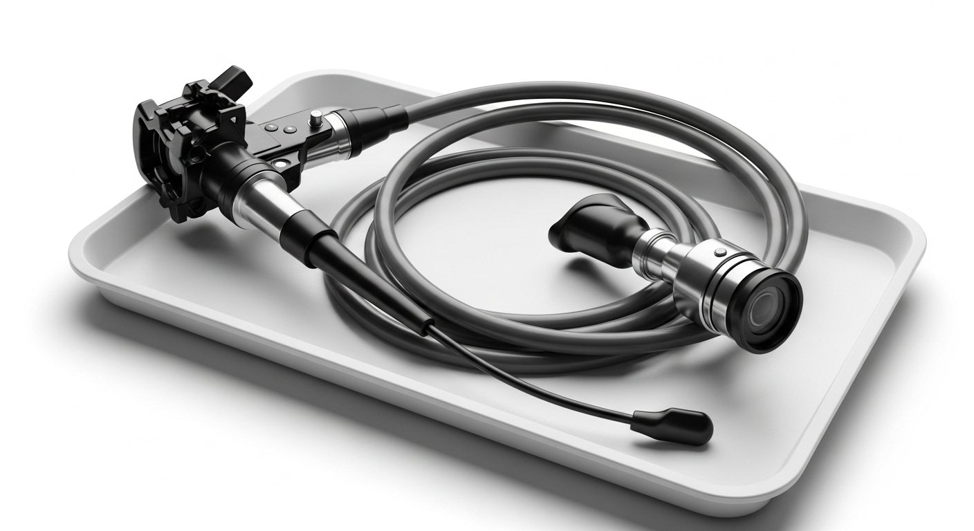
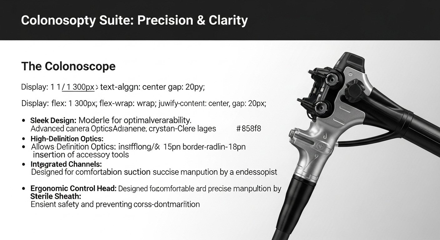
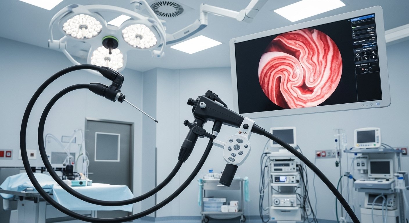
Current colonoscopes feature high-definition cameras, water jets for cleaning, and channels for inserting instruments. The scope’s flexibility allows navigation through the colon’s natural curves at the splenic and hepatic flexures. Carbon dioxide insufflation expands the colon for better visualization while reducing post-procedure discomfort compared to room air.
Gastroscopy Examination Areas
Gastroscopy begins with the endoscope passing through your mouth, down the esophagus, into the stomach, and reaching the duodenum’s first and second portions. The procedure examines the esophageal lining for inflammation or varices, the gastroesophageal junction for reflux damage, the stomach lining for ulcers or inflammation, and the duodenal walls for erosions or abnormalities.
The gastroscope’s smaller diameter (9-12mm) compared to the colonoscope (12-15mm) allows easier passage through the upper digestive tract. The procedure duration ranges from 5-15 minutes under conscious sedation. Narrow-band imaging technology enhances visualization of blood vessels and surface patterns, improving detection of subtle abnormalities.
Preparation Requirements
Colonoscopy Preparation
Bowel preparation begins 1-2 days before your colonoscopy with dietary modifications—avoiding seeds, nuts, and high-fiber foods. The day before the procedure, you’ll consume only clear liquids including water, clear broth, apple juice, and black coffee without milk.
The bowel cleansing solution, typically polyethylene glycol or sodium phosphate, requires drinking 2-4 liters split between evening and morning doses. The solution triggers frequent bowel movements over 2-4 hours, clearing stool from your colon. Some preparations include flavoring packets to improve palatability.
Medication adjustments may include stopping iron supplements 5 days prior, adjusting blood thinners according to your physician’s instructions, and continuing most other medications with small sips of water. Diabetic patients receive specific instructions for insulin or oral medication dosing during the fasting period.
Gastroscopy Preparation
Gastroscopy requires fasting for 8-12 hours before the procedure to ensure an empty stomach. Clear liquids may be permitted up to 2 hours before, depending on your anesthesiologist’s protocol. Unlike colonoscopy, no bowel preparation solution is needed.
Remove dentures, contact lenses, and jewelry before the procedure. Inform your doctor about dental crowns or loose teeth that might affect endoscope insertion. Patients with heart valve conditions may require antibiotic prophylaxis 30-60 minutes before the procedure.
Conditions Diagnosed
Colonoscopy Detects
Colorectal polyps appear as raised lesions on the intestinal wall, classified as adenomatous, hyperplastic, or sessile serrated based on histology. Adenomatous polyps carry malignant potential, with risk increasing for larger polyps. The adenoma detection rate serves as a quality indicator for colonoscopy.
Inflammatory bowel diseases manifest as continuous inflammation in ulcerative colitis or patchy inflammation in Crohn’s disease. Colonoscopy reveals mucosal friability, ulcerations, pseudopolyps, and strictures. Biopsy samples confirm diagnosis and assess disease severity.
Diverticular disease presents as outpouchings in the colon wall, most commonly in the sigmoid region. Colonoscopy identifies diverticula location, estimates burden, and excludes complications like bleeding or perforation. Colorectal cancer appears as irregular masses, strictures, or ulcerated lesions requiring biopsy.
Gastroscopy Detects
Peptic ulcers present as mucosal defects in the stomach or duodenum, with gastric ulcers typically along the lesser curvature and duodenal ulcers in the bulb. The endoscope assesses ulcer size, depth, and characteristics suggesting benign versus malignant etiology. Helicobacter pylori testing through rapid urease test or biopsy guides treatment decisions.
Gastroesophageal reflux disease causes esophageal mucosal changes graded using the Los Angeles Classification from Grade A (minimal breaks) to Grade D (circumferential breaks). Barrett’s esophagus, a complication of chronic reflux, shows salmon-colored mucosa replacing normal esophageal lining. Prague criteria document Barrett’s extent using circumferential and maximal measurements.
Esophageal varices, dilated veins from portal hypertension, require grading for bleeding risk assessment. Gastric cancer may appear as ulcerated masses, linitis plastica, or subtle mucosal irregularities. Early gastric cancer detection improves with chromoendoscopy and magnification techniques.
Sedation and Recovery
During the Procedures
Conscious sedation using midazolam and fentanyl provides comfort while maintaining protective reflexes. The anesthesiologist monitors oxygen saturation, heart rate, and blood pressure throughout. Propofol sedation provides deeper sedation with faster recovery.
Gastroscopy patients receive throat spray containing lidocaine to suppress the gag reflex. A mouth guard protects teeth and the endoscope during insertion. Colonoscopy patients lie on their left side initially, with position changes as needed for scope advancement.
💡 Did You Know?
Virtual reality headsets and music therapy may reduce anxiety and sedation requirements during endoscopic procedures, with some centers providing these complementary comfort measures.
Post-Procedure Recovery
Recovery room monitoring continues for 30-60 minutes until sedation effects diminish. Vital signs stabilize, and cognitive function returns gradually. Discharge requires a responsible adult escort, as driving remains prohibited for 24 hours post-sedation.
Colonoscopy recovery may include mild bloating from residual gas, resolving with walking and position changes. Dietary progression begins with light meals, advancing to regular diet as tolerated. Minor bleeding after polypectomy typically stops spontaneously.
Gastroscopy recovery involves throat numbness lasting 1-2 hours, requiring delayed eating and drinking to prevent aspiration. Mild throat discomfort resolves within 24-48 hours. Abdominal bloating from air insufflation improves with gentle movement.
Screening Guidelines and Timing
Colonoscopy Screening
Average-risk individuals begin colonoscopy screening at age 50, repeating every 10 years if normal. Earlier screening at age 40, or 10 years before the youngest affected relative’s diagnosis age, applies to those with family history of colorectal cancer.
High-risk groups requiring more frequent surveillance include patients with previous adenomatous polyps (repeat in 3-5 years depending on polyp characteristics), inflammatory bowel disease (annual or biannual after 8-10 years of disease), and hereditary cancer syndromes (annual screening from age 20-25).
⚠️ Note
Positive fecal immunochemical test results require colonoscopy within 8 weeks for cancer detection, as delays reduce screening program effectiveness.
Gastroscopy Screening
Routine gastroscopy screening lacks universal guidelines for average-risk populations. Specific groups benefit from surveillance including Barrett’s esophagus patients (every 3-5 years without dysplasia), individuals with family history of gastric cancer (discussion with gastroenterologist regarding timing), and those with hereditary diffuse gastric cancer syndrome (annual surveillance from age 20).
Symptomatic indications for gastroscopy include persistent dyspepsia in patients over 40, new-onset reflux symptoms unresponsive to treatment, dysphagia or odynophagia, unexplained iron deficiency anemia, and persistent vomiting.
Combined Procedures
Performing colonoscopy and gastroscopy during the same session offers several advantages:
- Single sedation exposure reduces cumulative risks and recovery time
- One preparation day minimizes work absence and caregiver burden
- Comprehensive evaluation addresses both upper and lower gastrointestinal symptoms simultaneously
The gastroscopy typically precedes colonoscopy to prevent contamination of the upper tract with colonic bacteria. Total procedure time extends to 35-60 minutes. Recovery protocols remain similar to single procedures, with dietary advancement following standard guidelines.
Discuss with a healthcare professional about scheduling both procedures if experiencing symptoms suggesting both upper and lower gastrointestinal pathology.
What Our Colorectal Surgeon Says
The decision between colonoscopy vs gastroscopy hinges on symptom localization and screening requirements. Epigastric pain, heartburn, and difficulty swallowing clearly indicate gastroscopy as the initial investigation.
Quality bowel preparation determines colonoscopy success more than any other modifiable factor. Split-dose preparation, completing the second dose 4-6 hours before the procedure, achieves optimal cleansing. Patients who cannot tolerate standard preparations have low-volume or tablet alternatives available.
Current endoscopic technology continues advancing detection capabilities. Artificial intelligence-assisted polyp detection, confocal laser endomicroscopy for real-time histology, and capsule endoscopy for small bowel evaluation expand diagnostic possibilities beyond traditional endoscopy.
Putting This Into Practice
- Schedule screening colonoscopy at age 50 if average-risk, earlier with family history or concerning symptoms
- Document family history of gastrointestinal cancers to guide appropriate screening intervals
- Maintain a symptom diary noting pain location, timing, and triggers to help your healthcare professional determine whether colonoscopy or gastroscopy is indicated
- Follow preparation instructions precisely, including dietary restrictions and medication timing
- Arrange transportation and post-procedure support before your appointment day
When to Seek Professional Help
- Blood in stool or black, tarry stools
- Persistent abdominal pain unrelieved by bowel movements
- Unexplained weight loss
- Changes in bowel habits lasting more than several weeks
- Difficulty swallowing or food sticking in throat/chest
- Persistent heartburn unresponsive to medication
- Chronic nausea or recurrent vomiting
- Iron deficiency anemia without obvious cause
- Positive fecal immunochemical or occult blood test
Commonly Asked Questions
Can I have both procedures done on the same day?
Yes, combined procedures occur frequently under single sedation. The gastroscopy precedes colonoscopy to maintain sterility. Total procedure time increases by 10-15 minutes compared to colonoscopy alone. Recovery follows standard protocols with no additional restrictions.
Which procedure is more uncomfortable?
Neither procedure causes significant discomfort with adequate sedation. Gastroscopy may trigger brief gagging during insertion, while colonoscopy can cause cramping during air insufflation. Most patients report minimal memory of either procedure due to sedation’s amnestic effects.
How accurate are these procedures for detecting cancer?
Colonoscopy can detect colorectal cancers and remove precancerous polyps during the same procedure. Gastroscopy identifies gastric and esophageal cancers, though early-stage lesions require enhanced imaging techniques. Biopsy confirmation provides definitive diagnosis.
What are the alternatives to these procedures?
CT colonography offers non-invasive colon imaging but cannot remove polyps or perform biopsies. Capsule endoscopy visualizes the small intestine but provides limited views of stomach and colon. Barium studies have largely been replaced by endoscopic evaluation.
How long should I wait between screening procedures?
Normal colonoscopy results permit 10-year screening intervals for average-risk individuals. Gastroscopy lacks defined screening intervals for asymptomatic patients. Surveillance intervals depend on initial findings—adenomatous polyps require 3-5 year follow-up, while Barrett’s esophagus needs evaluation every 3-5 years. A healthcare professional can provide guidance on appropriate screening schedules based on individual circumstances.
Conclusion
Colonoscopy screens for colorectal cancer and evaluates lower gastrointestinal symptoms, while gastroscopy investigates upper digestive complaints and complications. Your symptoms, risk factors, and screening timeline determine which procedure you need. Proper preparation significantly impacts procedure success and diagnostic accuracy. For a clearer understanding of what each test involves, read our colonoscopy guide and learn how it supports accurate diagnosis and effective treatment.
If you’re experiencing rectal bleeding, changes in bowel habits, persistent heartburn, or difficulty swallowing, consult with a colorectal specialist to determine the most appropriate endoscopic evaluation.